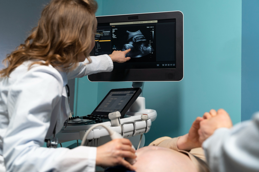
Ultrasound Imaging

Ultrasound Imaging is a non-invasive diagnostic tool widely used in obstetrics and gynecology to monitor the health of both the mother and baby throughout pregnancy. It uses high-frequency sound waves to create images of the internal organs, tissues, and developing fetus, providing real-time visual insights without the use of radiation.
In pregnancy, ultrasound imaging is essential for:
Fetal Development Monitoring: Ultrasound helps track the baby’s growth, detect heartbeat, and assess overall health. It also helps identify any congenital abnormalities early on.
Determining Gestational Age: An early ultrasound provides an accurate estimation of the baby’s age and due date.
Assessing Placental Position: Ultrasound checks for placental issues such as placenta previa, ensuring a safer delivery plan.
Detecting Multiple Pregnancies: It confirms whether a woman is carrying more than one baby, allowing for tailored care.
Guiding High-Risk Pregnancy Management: In cases of gestational diabetes or preeclampsia, regular ultrasounds monitor the baby’s condition to ensure timely interventions.
Examining Fetal Positioning: Helps determine the baby’s position, such as breech, guiding decisions about delivery.
Ultrasound is also used in gynecological care to diagnose conditions such as ovarian cysts, fibroids, and other abnormalities. Dr. S. Vidhyullatha ensures that her patients receive high-quality ultrasound imaging as part of comprehensive prenatal and gynecological care.


